Advantages of MRI in rheumatology: a detailed look

Contents
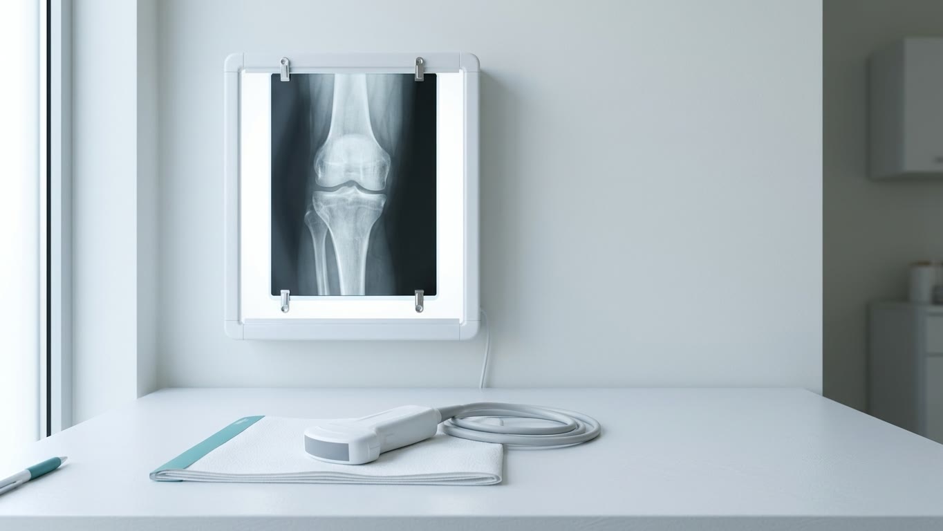
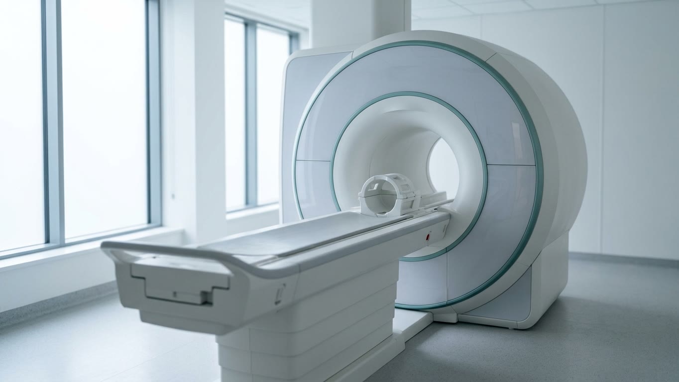
Magnetic resonance imaging (MRI) is the most sensitive imaging method in rheumatology. It shows inflammation and early damage where X-ray and ultrasound still see nothing. Let us look at what exactly MRI shows, when it is really needed and when it is not, whether contrast is required and how to prepare for the scan.
1. What MRI shows
- Synovitis and tenosynovitis — inflammation of the joint lining and tendon sheaths, fluid in the joint. Ultrasound sees this too, but MRI reaches deep joints and the spine, where ultrasound cannot.
- Bone marrow oedema (osteitis) — MRI’s main advantage. It cannot be seen on X-ray or ultrasound. In rheumatoid arthritis, osteitis is the strongest predictor of future bone erosions.
- Erosions — MRI and ultrasound find them earlier than X-ray, and MRI also shows whether the process around them is active.
- Sacroiliitis — inflammation of the sacroiliac joints in axial spondyloarthritis. MRI sees it months or years before changes appear on X-ray.
- Osteonecrosis — MRI detects it at an early stage, before the bone collapses.
Ultrasound has its own advantages: it is cheaper, more available, has no contraindications and lets the doctor watch a joint in motion. So the methods do not compete — they complement each other.
2. When a rheumatologist orders MRI
- Suspected axial spondyloarthritis — the most common reason. MRI of the sacroiliac joints is done when examination and X-ray are not enough for a diagnosis, and in young patients with recent back pain it is often done instead of an X-ray.
- Early rheumatoid arthritis or suspicious joint pain without obvious arthritis — MRI finds hidden inflammation.
- Suspected osteonecrosis — MRI is the method of choice.
- Assessing disease activity in selected cases — when it is important to know whether inflammation remains.
3. When MRI is not needed
- If changes are already clearly visible on X-ray, MRI adds nothing to the diagnosis.
- Repeating MRI “just in case” soon after a normal result is usually useless: with back pain, a repeat scan after 3 months to 2 years is positive in only 5–15% of patients.
- MRI is not used as a screening test for people without symptoms.
If you take anti-inflammatory drugs regularly, tell your doctor: they can reduce the signs of inflammation on the images.
4. Can MRI see the disease before symptoms?
Yes. In people with joint pain but no obvious arthritis yet, hidden inflammation on MRI appeared a few months before arthritis. If there was no inflammation on MRI, arthritis developed rarely — in about 6% of patients. With both inflammation on MRI and anti-citrullinated peptide antibodies (ACPA), arthritis developed within a year in 71%.
But bone marrow oedema also occurs in healthy people: after childbirth, in athletes, with mechanical strain. So an MRI finding is always judged together with symptoms, examination and blood tests, never on its own.
5. Is contrast needed?
For most rheumatology scans contrast is not needed. In spondyloarthritis, for example, inflammation is clearly visible on special sequences without contrast. It is used in selected cases, as the doctor decides.
If contrast is needed:
- Which agents. Contrast agents contain gadolinium. In 2017 the European Medicines Agency suspended several “linear” agents for intravenous use — among them Omniscan and Magnevist — because their gadolinium stays longer in the tissues, including the brain. No harm from this has been proven, but more stable “macrocyclic” agents are now preferred. You can ask the imaging centre which agent they use.
- Kidneys. Contrast is removed by the kidneys within a few hours. With severe kidney disease or on dialysis the safest agents are chosen — be sure to mention kidney disease in advance.
- Allergy. Reactions happen, but rarely. If you have reacted to contrast before, tell the doctor.
- Pregnancy. Contrast is avoided in pregnancy whenever possible.
6. Safety and contraindications
MRI does not use X-rays. The limitations come from the strong magnetic field:
- Pacemakers and other devices. Many modern pacemakers are MRI-compatible under certain conditions, but the decision is made only from the documents for the specific device.
- Metal implants. “Titanium is always safe” is an oversimplification. What matters is not the type of metal but the labelling of the specific implant: safe, conditional or unsafe. Bring the documents for any implants, clips or stents.
- Metal fragments, especially in the eye, must be reported in advance.
- Pregnancy. No harmful effect of MRI on the baby is known; the decision is made individually.
- Claustrophobia. A scanner with a wide, short tunnel or an open scanner may help. But open scanners usually give less detailed images — which matters when looking for small areas of inflammation and erosions.
7. How to prepare and how long it takes
- A joint MRI usually takes 20–45 minutes, depending on the area and whether contrast is used.
- Arrive about 30 minutes early: you will need to fill in a safety questionnaire.
- You can eat, drink and take your medicines as usual unless told otherwise.
- On the day, do not use hairspray, make-up, powder or creams: they may contain metal particles.
- Tell the staff about implants, operations, metal fragments, reactions to contrast and kidney disease.
- You need to lie still during the scan. The machine is noisy — earplugs or headphones are usually provided.
- If you are given a sedative for claustrophobia, someone else must drive you home.
8. MRI, X-ray and ultrasound compared
| Feature | MRI | X-ray | Ultrasound |
|---|---|---|---|
| Radiation | No | Yes, low | No |
| Bone marrow oedema | Yes | No | No |
| Synovitis | Yes | No | Yes |
| Deep joints and spine | Yes | Bones only | Poorly |
| Early erosions | Yes | Late | Yes |
| Availability and cost | More expensive | Widely available | Widely available |
Disclaimer: This article is for information only. Whether you need an MRI, with or without contrast, is decided by your doctor.
Sources
- Minopoulou I et al. Imaging in inflammatory arthritis: progress towards precision medicine. Nature Reviews Rheumatology, 2023.
- Navarro-Compán V et al. Axial Spondyloarthritis. Lancet, 2025.
- De Craemer AS, Łukasik Z, Carron P. Use of Imaging in Axial Spondyloarthritis for Diagnosis and Assessment of Disease Remission in the Year 2022. Current Rheumatology Reports, 2022.
- Bakker PAC et al. Is it Useful to Repeat MRI of the Sacroiliac Joints After Three Months or One Year in Patients With Suspected Axial Spondyloarthritis? Arthritis & Rheumatology, 2019.
- van Steenbergen HW et al. Clinical Factors, Anticitrullinated Peptide Antibodies and MRI-detected Subclinical Inflammation in Relation to Progression From Clinically Suspect Arthralgia to Arthritis. Annals of the Rheumatic Diseases, 2016.
- Runge VM. Gadolinium Deposition After Injections of Gadolinium-Based Contrast Agents and the EMA Recommendation for Suspension of 4 Linear Agents. Investigative Radiology, 2017.
- ACR Manual on Contrast Media. American College of Radiology, 2025.
- ACR Manual on MR Safety. American College of Radiology, 2024.
Recognise your symptoms?
An article does not replace an in-person consultation. Get in touch to book an appointment — we will go through your situation in person. Consultations are held in Russian.
Get in touch to book