Comparing imaging methods in rheumatology: ultrasound, X-ray, MRI

Contents
Rheumatology has three main imaging methods — ultrasound, X-ray and MRI. They do not replace but complement each other: each answers its own question best. Let us look at what each method sees, where doctors usually start and which common beliefs about them are wrong.
1. Joint ultrasound
What it sees well:
- inflammation of the joint lining (synovitis) and fluid in the joint; in Doppler mode, active inflammation with increased blood flow;
- tendons and their sheaths, tears, bursae (bursitis);
- the places where tendons attach to bone (enthesitis), including hidden inflammation;
- the bone surface and erosions — in early rheumatoid arthritis ultrasound finds them more often than X-ray;
- crystal deposits in gout (the “double contour” sign, tophi) and in calcium pyrophosphate disease.
Limitations:
- ultrasound does not pass through bone, so it cannot see the bone marrow or marrow oedema;
- it sees deep joints poorly, including the sacroiliac joints;
- the result depends heavily on the doctor’s experience.
Advantages: no radiation and no contraindications; many joints can be checked at once, and a joint can be seen in motion. Under ultrasound guidance, aspirations and injections into joints, tendon sheaths and bursae are more accurate and safer.
2. X-ray
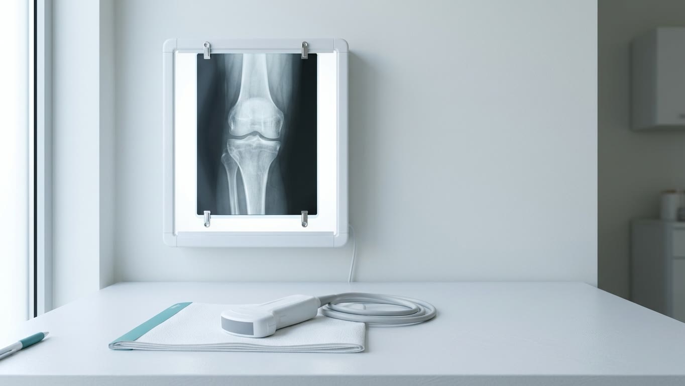
What it sees well: bones. It is the standard for diagnosing osteoarthritis — narrowing of the joint space, osteophytes (bony growths), hardening of the bone under the cartilage. X-ray also shows established damage: erosions in rheumatoid arthritis and gout, changes in the sacroiliac joints in spondyloarthritis.
Limitations: it does not show the joint lining, cartilage, ligaments or tendons. In early arthritis the image is usually normal — erosions and changes appear after months or years.
Advantages: quick, cheap and available almost everywhere.
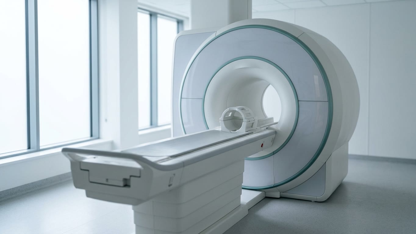
3. MRI
What it sees well: every structure of the joint, including deep joints and the spine. Its main advantage is bone marrow oedema (osteitis), which neither ultrasound nor X-ray can see. In rheumatoid arthritis it predicts future erosions, and in spondyloarthritis MRI finds inflammation of the sacroiliac joints years earlier than X-ray.
Limitations: more expensive, less available, takes longer, and has contraindications (some pacemakers and implants). More in the article “Advantages of MRI in rheumatology”.
4. What each method sees
| What we look for | X-ray | Ultrasound | MRI |
|---|---|---|---|
| Synovitis | No | Yes | Yes |
| Early erosions | Late | Yes | Yes |
| Bone marrow oedema | No | No | Yes |
| Osteoarthritis | Yes, the standard | Partly | Yes, but not first |
| Tendons and bursae | No | Yes | Yes |
| Crystals in gout | Late | Yes | Tophi only |
| Sacroiliac joints | Late changes | Poorly | Yes, early |
5. Where doctors usually start
- Suspected inflammatory arthritis — ultrasound or MRI: they find inflammation earlier than X-ray. For the joints of the hands and feet, ultrasound is usually first.
- Knee pain in an older person — an X-ray taken standing up: the standard for osteoarthritis. Ultrasound is added to assess fluid, inflammation or crystals.
- Suspected gout — ultrasound can show crystal deposits. But a normal ultrasound does not rule out early gout, and the most accurate method is joint fluid analysis.
- Back pain with suspected spondyloarthritis — an X-ray of the sacroiliac joints or MRI. In young patients with recent pain, MRI is preferred.
- Shoulder pain — ultrasound: tendons, tears and bursae are clearly visible. MRI is for complex cases and before surgery.
6. How long it takes and how much it costs
- X-ray — a few minutes, inexpensive, available almost everywhere.
- Ultrasound — usually 10–30 minutes, cheaper and more available than MRI.
- MRI — usually 30–45 minutes or longer, the most expensive of the three.
7. Myths about imaging
- “Ultrasound cannot see bone.” Not quite. Ultrasound sees the bone surface and erosions on it well, but not what is inside the bone.
- “MRI is always better.” No. For osteoarthritis X-ray is the standard; for shoulder tendons and injections ultrasound is often more convenient. The best method is the one that answers the doctor’s question.
- “X-ray does not show early arthritis.” True: at the start of the disease the image is usually normal. That is why ultrasound and MRI are chosen for early diagnosis.
- “X-ray is dangerous.” The dose from a joint X-ray is very small — roughly the same as a few days of natural background radiation. But every X-ray should be justified.
8. What about osteoporosis?
X-ray, ultrasound and MRI do not measure bone density. There is a separate method for that — densitometry (DXA). It measures bone mineral density in the spine, hip and forearm, and osteoporosis is diagnosed from it. The radiation dose from DXA is comparable to 1–2 days of natural background radiation.
Disclaimer: This article is for information only. Which test you need is decided by your doctor.
Sources
- Minopoulou I et al. Imaging in inflammatory arthritis: progress towards precision medicine. Nature Reviews Rheumatology, 2023.
- Filippucci E, Di Geso L, Grassi W. Progress in imaging in rheumatology. Nature Reviews Rheumatology, 2014.
- Kaeley GS, Bakewell C, Deodhar A. The importance of ultrasound in identifying and differentiating patients with early inflammatory arthritis. Arthritis Research & Therapy, 2020.
- Gessl I et al. Systematic Literature Review to Inform the EULAR Recommendations for the Use of Imaging in Crystal-Induced Arthropathies. Annals of the Rheumatic Diseases, 2024.
- Taljanovic MS et al. High-Resolution US of Rheumatologic Diseases. RadioGraphics, 2015.
- Bittar M, Deodhar A. Axial Spondyloarthritis: A Review. JAMA, 2025.
- Fukui S et al. Current Evidence and Practical Knowledge for Ultrasound-Guided Procedures in Rheumatology. Best Practice & Research Clinical Rheumatology, 2023.
- Link TM, Kazakia G. Update on Imaging-Based Measurement of Bone Mineral Density and Quality. Current Rheumatology Reports, 2020.
Recognise your symptoms?
An article does not replace an in-person consultation. Get in touch to book an appointment — we will go through your situation in person. Consultations are held in Russian.
Get in touch to book